
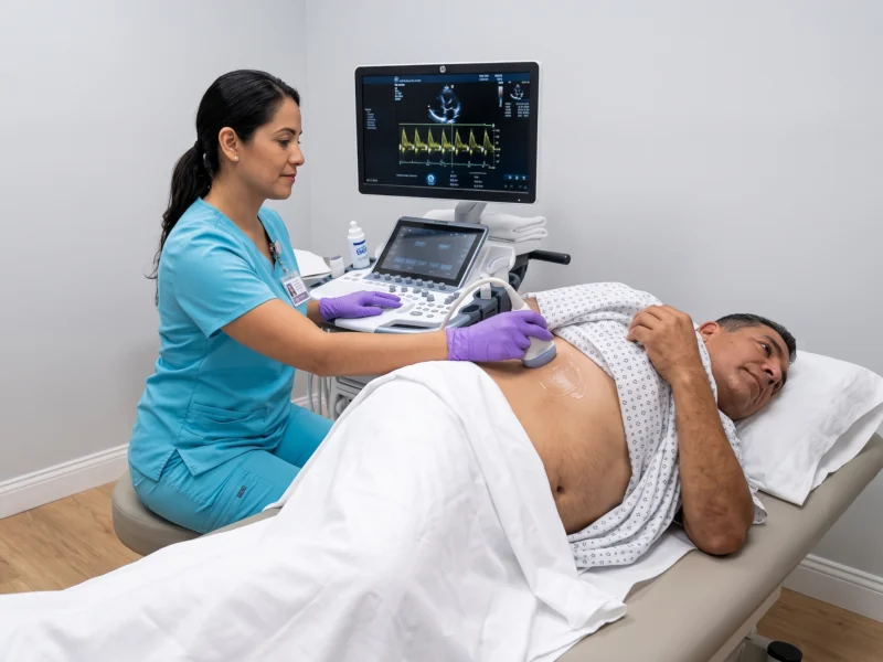
Echocardiography (ECHO) serves as the definitive diagnostic ultrasound modality for capturing dynamic, real-time visualizations of cardiac architecture and mechanics. Utilizing high-frequency sonic waves entirely free of ionizing radiation, this non-invasive examination delivers meticulous anatomical clarity across cardiac chambers, myocardial wall kinetics, and valvular hemodynamics to accurately assess complete cardiovascular performance.
Equipped with advanced cardiac Doppler capabilities, our diagnostic suites evaluate blood flow velocity and identify structural anomalies, valvular regurgitation, or stenosis with exceptional precision. Administered by experienced cardiac sonographers, every evaluation equips referring clinicians and cardiologists with essential objective metrics, including left ventricular ejection fraction and chamber dimensions, facilitating timely and informed therapeutic decisions.
Primary Clinical Applications
Heart Valve Evaluation: Detects valve narrowing (stenosis), leakage (regurgitation), or prolapse in the mitral, aortic, tricuspid, and pulmonary valves.
Assessment of Cardiac Function: Quantifies ejection fraction (EF) to determine how effectively the heart pumps oxygenated blood throughout the body.
Heart Failure & Cardiomyopathy: Identifies ventricular hypertrophy (thickened muscle), chamber enlargement, and chronic cardiac strain.
Investigation of Symptoms: Evaluates the clinical origin of unexplained shortness of breath, heart murmurs, peripheral edema (leg swelling), or chest discomfort.
Pericardial Disease: Screens for fluid accumulation around the heart (pericardial effusion) or inflammatory pericardial thickening.
Patient Instructions & Exam Preparation
No fasting required: You may eat, drink water, and take all your daily prescribed heart and blood pressure medications as scheduled.
Clothing: Wear a comfortable two-piece outfit or a button-down shirt to allow easy access to the chest area.
Skin preparation: Do not apply body lotions, oils, or powders to the chest on the day of the exam to maintain clear acoustic transducer contact.
Comfort & sensation: The procedure is entirely painless and radiation-free; a warm, hypoallergenic acoustic gel is applied to the chest while you lie comfortably on your left side.
Exam duration: The examination typically takes 30 to 45 minutes, with comprehensive diagnostic reports delivered promptly to your ordering physician.